Overview
What is a USG WHOLE ABDOMEN ?
A USG Whole Abdomen, also known as an Ultrasound of the Whole Abdomen, is a non-invasive diagnostic imaging test that uses high-frequency sound waves to create real-time images of the organs and structures located within the abdominal cavity.
It is one of the most commonly performed and safest imaging tests in medicine. The test helps doctors visualize, study, and evaluate the condition of vital internal organs without using radiation, injections, or invasive instruments.
It Called “Whole Abdomen Because the test covers both the upper and lower abdomen, including organs of the digestive system, urinary system, and reproductive organs (in women). It’s more extensive than a “USG Upper Abdomen,” which focuses only on the liver, gallbladder, pancreas, spleen, and kidneys.
What does a USG WHOLE ABDOMEN diagnose?
A USG Whole Abdomen helps doctors see what’s happening inside your stomach area and detect any problems in your internal organs — like the liver, kidneys, gallbladder, pancreas, spleen, urinary bladder, and reproductive organs.
It can identify a wide range of conditions that may cause pain, swelling, or other symptoms.
1. Liver Problems
- Fatty liver (too much fat stored in the liver)
- Hepatitis (liver inflammation or infection)
- Cirrhosis (long-term liver damage)
- Liver cysts or tumors
- Gallbladder and Bile Duct Issues
- Gallstones (stones that form in the gallbladder)
- Cholecystitis (infection or swelling of the gallbladder)
- Bile duct blockage
- Kidney and Urinary Tract Conditions
- Kidney stones
- Kidney cysts or swelling
- Urinary tract infections (UTI)
- Bladder stones or wall thickening
- Pancreatic Disorders
- Pancreatitis (inflammation of the pancreas)
- Pancreatic cysts or growths
- Spleen Problems
- Enlarged spleen (seen in infections or blood disorders)
- Injury or rupture of the spleen
- Abdominal Fluid or Masses
- Ascites (extra fluid in the abdomen)
- Abnormal lumps, cysts, or tumors
- Reproductive Organ Issues (checked if included in the scan)
- In women: Ovarian cysts, uterine fibroids, or pelvic infections
- In men: Enlarged prostate or prostate infections
- Blood Vessel Problems
- Abnormal blood flow in the aorta or major abdominal vessels
- Aneurysms (bulging of blood vessel walls)
Indications
- Abdominal pain: Persistent or severe pain in the belly, particularly if the cause is unclear.
- Abdominal swelling or bloating: Unexplained enlargement of the abdomen that may indicate a problem with the liver, kidneys, or the presence of abnormal fluid (ascites).
- Abnormal liver or kidney function tests: Blood test results indicating that these organs may not be functioning correctly.
- Nausea and vomiting: Persistent or recurring episodes without a known cause.
- Jaundice: Yellowing of the skin and eyes, which can suggest liver or gallbladder issues.
- Changes in bowel habits: Persistent diarrhea, constipation, or other intestinal issues.
- A palpable mass or lump: When a doctor can feel an abnormal lump during a physical examination.
- Painful urination or blood in urine: Symptoms that may indicate kidney stones, a kidney infection, or a bladder problem.
- Fever of unknown origin: A fever that cannot be explained by other conditions could point to an abdominal infection, such as an abscess.
Diagnosing and monitoring specific conditions
- Gallstones or kidney stones: An ultrasound is highly effective at visualizing and detecting the presence of stones.
- Liver disease: Conditions like fatty liver disease, hepatitis, or cirrhosis can be diagnosed by assessing the liver’s size, texture, and other features.
- Pancreatic issues: Used to check for inflammation (pancreatitis), cysts, or tumors.
- Enlarged spleen (splenomegaly): The test can confirm if the spleen is enlarged, which is a symptom of many underlying conditions.
- Abdominal aortic aneurysm (AAA): An ultrasound is often used as a screening tool to check for an enlargement of the abdominal aorta.
- Tumors, cysts, or abscesses: To identify and evaluate any abnormal masses in the abdominal organs.
- Appendicitis: Can be used to help confirm the diagnosis of a suspected inflamed appendix.
Test Details
Who Performs a USG Whole Abdomen?
A USG Whole Abdomen is done by a radiologist or a trained sonographer (ultrasound technician).
They use a special handheld device to take pictures of your internal organs.
The radiologist then studies these images and prepares a report for your doctor.
What Types of Scanners Are Used for a USG Whole Abdomen?
Types of Scanners Used for a USG Whole Abdomen
A USG Whole Abdomen can be performed using different types of ultrasound scanners depending on the level of detail required and the diagnostic purpose:
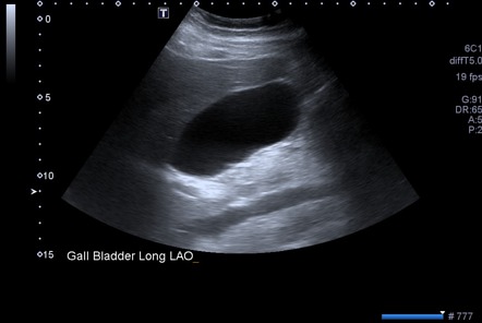
- 2D Ultrasound Scanner
- The most common type of scanner.
- Produces real-time black-and-white images of your organs.
- Helps check the size, shape, and basic structure of the liver, kidneys, gallbladder, spleen, pancreas, and bladder.
- 3D / 4D Ultrasound Scanner
- Produces three-dimensional images (3D) or live-motion images (4D).
- Gives a more detailed and clear view of organs and abdominal structures.
- Doppler Ultrasound Scanner
- Measures blood flow in abdominal vessels.
- Helps detect blockages, reduced blood flow, or aneurysms in major vessels like the aorta.
What happens before a USG WHOLE ABDOMEN?
Before a USG Whole Abdomen Preparing for the scan helps get clear and accurate images of your internal organs.
- Fasting:
- Usually, you may need to avoid food and drinks for 6–8 hours before the test.
- This is important to get a clear view of the liver, gallbladder, and pancreas.
- Full Bladder (if needed)
- For women, if pelvic organs like the uterus or ovaries are being checked, you may be asked to drink water and keep your bladder full.
- Comfortable Clothing
- Wear loose, comfortable clothes.
- You may be asked to remove belts, jewelry, or any metal objects from your abdomen.
- Medical History
- Inform the technician or doctor about any previous surgeries, medications, or medical conditions.
What happens during a USG WHOLE ABDOMEN ?
For a whole abdomen ultrasound (USG), the procedure is simple, quick, and painless. A technician, called a sonographer, will perform the scan by using a handheld device on your belly to create images of your internal organs. The entire process typically takes less than 30 minutes.
What happens during the exam
Get ready: You will be asked to lie down on your back on a comfortable examination table. You may also be asked to change into a hospital gown.
Gel application: The sonographer will apply a clear, water-based gel to your abdomen. The gel may feel a little cool and wet at first. This gel is essential for the sound waves to travel smoothly from the device into your body, eliminating any air pockets that could blur the images.
Scanning: The sonographer will press a handheld tool called a transducer against your skin and move it around your abdomen. You may feel gentle pressure as they move the probe to get clear pictures of different organs.
Special instructions: During the scan, you may be asked to hold your breath for a few seconds. This helps get a better view of certain organs, like the liver. You might also be asked to turn onto your side.
Listen for blood flow (if needed): If your doctor requested a Doppler ultrasound, you might hear “whooshing” or pulsing sounds. This is normal and is just the machine measuring blood flow through your vessels.
Finishing up: Once the sonographer has captured all the necessary images, they will wipe the gel off your skin. The gel will not stain your clothes.
How Long Does a USG Whole Abdomen Take?
A USG Whole Abdomen usually takes about 20 to 30 minutes.
The exact time may vary depending on the organs being examined and if additional images or Doppler studies are needed.
The procedure is quick, painless, and safe, with no recovery time required.
The procedure is quick, painless, and safe, with no recovery time required.
What Happens After a USG Whole Abdomen ?
After your USG Whole Abdomen is completed, the next steps are simple and safe. The scan itself is non-invasive, painless, and requires no recovery time, so you can continue with your daily activities immediately.
- Review of Images
- The radiologist examines the ultrasound images carefully.
- They look at the size, shape, texture, and condition of all abdominal organs such as the liver, kidneys, gallbladder, pancreas, spleen, and bladder.
- If Doppler imaging was done, blood flow in the abdominal vessels is also evaluated.
- Preparing the Report
- Based on the findings, the radiologist prepares a detailed report.
- The report includes any abnormalities, organ enlargement, stones, cysts, fluid accumulation, or other issues detected during the scan.
- It also mentions if everything appears normal.
- Sharing Results with Your Doctor
- The report is sent to your referring doctor, who will review it with you.
Your doctor may explain the results, reassure you if everything is normal, or suggest further tests or treatment if any abnormality is found.
- No Special Care Needed
- There are no restrictions on eating, drinking, or activities after the scan.
- You do not need any rest or medication because the procedure is completely safe.
What are the benefits of a USG WHOLE ABDOMEN ?
- Non-invasive and Painless
- The test does not involve any cuts, injections, or radiation.
- You won’t feel pain, making it suitable for patients of all ages, including children and elderly people.
- Quick and Convenient
- The scan usually takes only 20–30 minutes.
- There’s no recovery time, and you can resume your daily activities immediately.
- Safe for Everyone
- Uses sound waves, not X-rays, so it is completely safe, even for pregnant women.
- Early Detection of Problems
- Helps identify issues in liver, kidneys, gallbladder, pancreas, spleen, and bladder before they become severe.
- Can detect stones, cysts, tumors, fluid accumulation, or organ enlargement early.
- Monitors Existing Conditions
- Doctors can track the progress of chronic conditions like fatty liver, kidney disease, or gallbladder problems.
- Helps in adjusting treatment plans based on real-time imaging.
- Guides Treatment Decisions
- Provides accurate images for doctors to plan surgery, medication, or other treatments.
- Reduces the need for more invasive procedures in many cases.
- Comprehensive Overview
- Examines all major abdominal organs in one scan, giving a complete picture of your abdominal health.
Risks of a USG Whole Abdomen
The USG Whole Abdomen is considered one of the safest medical imaging tests. It uses sound waves instead of radiation, so it’s generally risk-free.
Key Points About Risks
- No Radiation: Unlike X-rays or CT scans, ultrasound does not expose you to harmful radiation.
- Non-invasive: There are no injections or cuts, so infection or injury risk is extremely low.
- Safe for Everyone: It can be done safely in pregnant women, children, and elderly patients.
- Mild Discomfort Only: Some patients may feel slight pressure when the technician presses the transducer on the abdomen, but it does not cause pain.
Very Rare Risk
- Rarely, some people may have a minor allergic reaction to the gel, but this is extremely uncommon.
- No long-term side effects are associated with ultrasound scanning
A Note from North City Diagnostic The uncertainty of not knowing what’s happening inside your body can be stressful. A USG Whole Abdomen can provide clarity. This safe and non-invasive ultrasound test helps your healthcare provider detect problems like liver disease, kidney or gallbladder stones, pancreas or spleen issues, and fluid accumulation in the abdomen. Different types of ultrasound scanners are available depending on your needs and the level of detail required. Your provider will discuss the next steps based on your test findings.
Care at North City Diagnostic
- If you have digestive system concerns or abdominal discomfort, you need a team of experts you can trust. Our radiologists and trained sonographers at North City Diagnostic ensure accurate imaging and detailed reporting to help guide your doctor in your care plan.